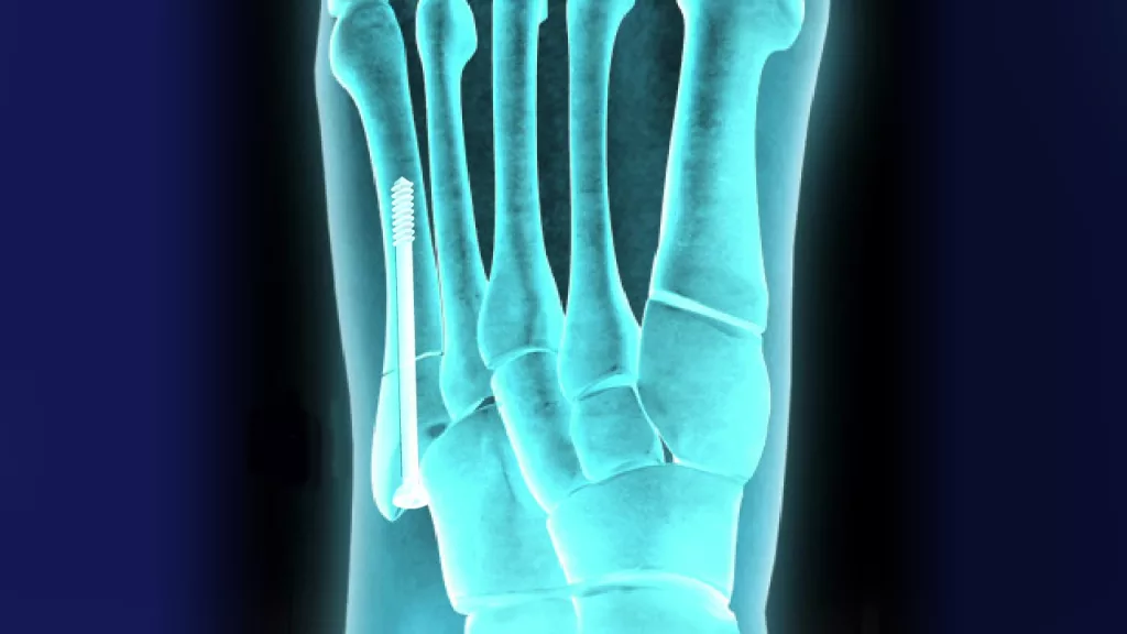
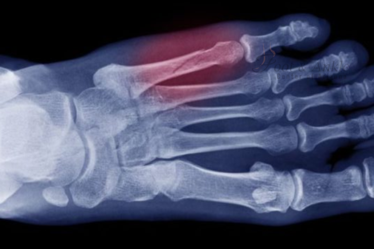
The Jones fracture is a selected type of fracture occurring at the metaphyseal-diaphyseal junction of the fifth metatarsal bone on the lateral side of the foot. The fracture is named after Sir Robert Jones, who first described it, and it is common among athletes and those with high-impact sports. This fracture is unusual or irritating because the nature of this fracture is a region of poor healing due to its location.
While not all fifth metatarsal fractures require surgery, Jones fractures often need one since the limited blood supply to this area affects natural healing. When conservative treatment, immobilization, and rest are inadequate for complete recovery, it is necessary to have a surgical operation, known as Jones fracture fixation, particularly open reduction and internal fixation.
Understanding Jones Fracture
A Jones fracture is a type of fracture that occurs near the base of the fifth metatarsal, where the wide base of the bone becomes the slender shaft. This junction area easily gets into trouble, especially in those whose feet are subjected to repeated stress, such as joggers, jumpers, and dancers, specifically those who pivot on one foot. The fracture can be due to sudden twisting of the foot or direct violence, while often playing basketball, soccer, or tennis.
The symptoms of a Jones fracture include swelling, pain on the outside of the foot, and inability to bear weight. Poor blood supply in this area can make a Jones fracture heal very slowly even with nonsurgical treatments like casting or immobilization. Some patients can manage with non-surgical measures; some people, either because of the severity or displacement of the fracture, may be advised that surgery would provide the most predictable means for the complete healing and return of function in the foot.
When Surgery is Required
Whether or not to resort to surgery depends on several factors, including the severity of the fracture the level of activity for the patient, and how well the bone is setting without any intervention. In the event of a displaced fracture or a highly active patient desiring a rapid return to sports participation, Jones fracture fixation surgery is usually indicated. That way, it ensures that the bone heals properly and minimizes the risk of serious long-term consequences, such as chronic pain or recurring fractures.
Procedure: Open Reduction and Internal Fixation (ORIF)
This surgery generally involves open reduction and internal fixation, a standard technique of fixation of many fractures whereby the bone fragments are restored to their normal position and held in place with hardware such as screws or plates. Here is a detailed look at the steps involved:
Preparation
The operation starts by positioning the patient appropriately, usually lying on the back and elevating the injured foot. Anesthesia keeps the patient comfortable without pain throughout the operation. It may be general anesthesia, in which case the patient sleeps; it may be regional anesthesia, where the leg and foot remain numb while the patient stays awake.
Incision and Access to Fracture
A small incision is made along the foot’s outer side to expose the fractured area of the fifth metatarsal. This allows direct access to the fractured bone so your surgeon can accurately align the fragments.
Reduction of the Fracture
When the fracture has become visible, the surgeon carefully moves the fragments around to restore their alignment. This step is called reduction and forms an important part of the operation because it allows the bone to heal in its correct anatomical position.
Internal Fixation with Screws or Plates
After the surgeon has aligned the bone, one or more screws are crossed through the fracture to stabilize the bone. Sometimes, other hardware such as plates may also be used for extra stability. These screws hold the ends of the fracture together, squeezing the bone fragments and hence speeding up the healing by minimizing the amount of movement occurring at the site of the fracture. The size of the fracture determines the size of the hardware, along with the needs of the patient.
Bone Grafting-if required
In some instances but rarely, especially in fractures that are slow to heal or bone loss, the surgeon may consider performing a bone graft. In this case, he may harvest bone from some other part of the body of the same patient or use some synthetic graft to stimulate his healing process. If the bone grafting is considered necessary, another incision may be performed to harvest the bone.
Closure of Incision
Following the reduction of the fracture and after the hardware is in place, the surgeon then closes the skin with sutures or staples. A sterile dressing is applied afterward, along with a splint, to protect the surgical site and to keep the foot below the knee immobilized.
Post-Surgical Care and Recovery
Generally, it is advised for a patient post-operatively not to bear weight on the involved foot for the initial seven to fourteen days. The foot remains in a splint or a cast at this period. For mobility assistance, crutches or a walker can be used, while ensuring that there will be no pressure on the foot. The goal of the initial weeks of rest is to allow the bone to begin to heal without the additional stress created by bearing weight.
Some surgeons may allow partial weight-bearing during the recovery process, and this approach is usually debated because of the risk of compromising the healing process. Generally speaking, most patients are told to place no weight on the foot whatsoever until a follow-up X-ray confirms that the bone is healing correctly.
A walking boot or brace can be initiated at about six weeks after surgery to transition the patient back to normal activity. The supportive device allows for progressive weight bearing while continuing the protection of the healing bone. Often during this period, physical therapy can be implemented to regain strength, flexibility, and balance of the foot.
Long-Term Recovery
Recovery from the Jones fracture fixation generally takes four months, though this may vary depending on the patient’s condition and the severity of the injury. After a few months, the patients will be able to resume light work; for high-impact sports and other such heavy activities, they may have to wait for the time the bone takes to heal.
One of the big positives regarding surgical intervention is that it can offer a more predictable and often faster recovery compared to nonsurgical treatments. By stabilizing the bone with screws or other hardware, the risk of non-union-poor or incomplete healing is reduced, and the patient can return to normal activity with a little more confidence.
Potential Risks and Complications
Infection, bleeding, nerve damage, and complications associated with anesthesia may also occur with Jones fracture fixation, as it is with any other surgery. In rare instances, the hardware used to fix the fracture may irritate or be uncomfortable enough to warrant a second surgery to remove it.
Other possible complications include non-union, a case where, even after surgery, the bone does not heal. This may be due to early return to activity by the patient or inadequate blood supply at the site of fracture. Close follow-up with the operating surgeon and adherence to post-operative care instructions are of utmost importance in minimizing these risks.
Conclusion
Jones fracture fixation surgery is safe and effective for fifth metatarsal fractures, especially among active patients who desire a quick return to full activities. Surgery to stabilize the bone accelerates the healing process more predictably and thus allows the full function of the foot to be restored with fewer complications.


