WBCT Unveils Rotational Dynamics in Ankle Joints
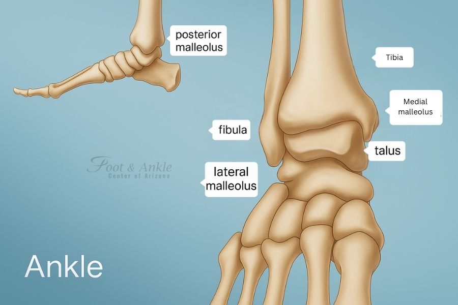
Weight-Bearing Computed Tomography and Rotational Dynamics of the Normal Distal Tibiofibular Joint. Little is known about the normal rotational dynamics of the distal tibiofibular (ankle) joint in upright weight-bearing conditions. Researchers conducted a study to assess these dynamics on weight-bearing cone beam computed tomography (WBCT), under physiological conditions. Thirty-two patients received low-dose WBCT scans, and the normal intersubject and intrasubject variation in neutral position and changes in maximal internal and external rotation of the ankle were studied.
Measurements included views from side to side (sagittal) translation of the fibula, and front to back (anterior and posterior) widths of the distal tibiofibular syndesmosis, tibiofibular clear space (TFCS) and rotation of the fibula (smaller the 2 shin bones). The fibula was located toward the front (anteriorly) in the tibial incisura in 88 percent of patients. When rotated, mean anteroposterior motion was 1.5 mm and mean rotation of the fibula was three degrees. No significant change existed in TFCS between internal and external rotation. The researchers concluded that the internal control of the contralateral ankle seemed to be a better reference than the population-based normal values.
Clinical Relevance: The current study provides the reference values to evaluate the rotational dynamics of a normal distal tibiofibular joint.
http://fai.sagepub.com/content/early/2016/02/25/1071100716634757.abstract


